Introduction
Diffraction is one of the most frequently encountered physical phenomena in the human world. It is responsible, among other things, for us being able to shout (and hear) ‘around’ a corner and explains why radio and television signals reach every hook, nook and cranny of a city. Diffraction is also the underlying mechanism of the iridescent colours of soap bubbles and petrol on a puddle, as well as the rainbow effect of so-called 'Newton's rings'.
Teachers the world over, together with backyard scientists, have demonstrated the occurrence of diffraction using one-slit and two-slit diffraction, diffraction about a thin wire or through a pin-point hole, using cheap lasers in the VIS (visible) part of the electromagnetic (EM) spectrum.
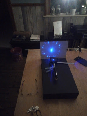
Here’s one of my one-slit experiments using a green pen-style laser (forgive the crappy wall paper used as background):

The laser's green light's wavelength here was 532 nm and the slit width about 30 micron (in the order of magnitude of a human hair)
The lighter areas show up where positive interference occurs, the darker ones where negative interference is found.
X-ray diffraction (XRD) – Bragg diffraction
Scientifically XRD has been used to probe the internal, atomic structure of materials like NaCl (table salt), crystalline DNA and minerals to name but a very, very few. The X-ray part of the EM stretches from wavelengths of 10 pm (‘hard’ X-rays) to 10 nm (‘soft’) X-rays). This is highly relevant because as an order of magnitude this corresponds approximately with significant length scales inside of said crystalline materials.
XRD basically works like this:

The horizontal black lines represent crystal planes (linear planar alignments of actual atoms inside the crystal under investigation). The inclined blue lines represent incident and diffracted X-rays, and α and β the angles of incidence and diffraction, respectively. It can then be shown that the formula at the bottom of the figure correctly predicts the values of α and β for which positive interference occurs. This manifests itself in a lighter area in the ‘detector’, e.g. a camera.
In the formula, aka the Bragg Condition, λ is the wavelength of the monochromatic) X-rays, d is the distance between the atomic planes in the crystal and n is an integer (1,2,3…, aka the 'order' of diffraction)
Clearly, with a suitable experimental set up, this opens up the exciting possibility of determining d (QED the case of DNA, Watson and Crick) and thus the crystalline structure of a material.
Note that in a real crystal, there are many possible sets of atomic planes where diffraction can occur, leading to complex diffraction patterns that can only be 'disentangled' by means of advanced math. Again, QED the case of DNA. Below a representation of a few planes (thick blue lines) in a 2 D cubic lattice (thin black lines, the nodes are atoms):

This type of so-called X-ray diffraction crystallography was discovered by a father and son team by the name of Bragg (in 1913)
Bragg diffraction using VIS light?
Would it be possible to apply the Bragg principle using VIS EM, using cheap pen-type lasers? In principle ‘yes’ but the Bragg condition shows such a material would have to possess “d-values” in the order of magnitude of the wavelength of the visible light which is approximately 400 to 800 nm (much larger than X-rays’ 10 pm to 10 nm range)
a. Construction of a 'pseudo-crystal' lattice using thin and transparent films:
Using various film substrates like cellophane, polypropylene/polyethylene, kitchen cling film and microscope slides, various ‘stacks’ were constructed and tested for diffraction using the red laser. While weak diffraction was obtained, it was not measurable. The thinnest film was about 50000 nm thick and most films weren’t transparent enough when stacked to about 0.5 mm thicknesses.
b. Iridescent ‘Merry Christmas’ banner:
Next up, a suggestion by ‘farcher’ at the physics.stackexchange.com/ website (of which I am a proud member): ‘Merry Christmas’ style banner film. Below a photo of a piece of film I cut off:

The material shows strong surface (reflective) VIS diffraction, which produces that very attractive shimmering colours effect. This material looks very different depending of the angle of visible light incidence.
The film is very, very thin and somewhat see through.
This led to the first successful VIS Bragg diffraction experiment of mine, using a purple laser of 432 nm wavelength, by shining it through the film and projecting the result onto a white paper 'detector':

The diffraction pattern, the dozen or so points arranged on a circle (approximately), are positive interference points, resulting from diffraction by internal atomic/molecular planes.
But the material is highly anisotropic and moving the incident laser a little, changes or destroys the observed pattern. This makes it unsuitable for any quantitative analysis. But we can be sure the “d values” are in the order of magnitude of 400 to 800 nm.
The diffraction here is caused by superthin 'confetti', dispersed throughout the resin matrix of the film. Varying orientations of the confetti then explains the unusual iridescence.
c. Synthetic Opal as a ’pseudo-crystal':
Here is a stock photo of a synthetic Opal cube, which I then purchased:

The iridescent colours of white (non-monochromatic) light is caused by Bragg-style diffraction (it is a truly beautiful - and cheap - object) I believe my purchase is a so-called Gilson-type synthetic Opal.
A simple experiment using the red (obviously monochromatic) laser showed very strong, consistent and clear diffraction. It’s so evident that the cube could be used as a beam splitter: much of the laser beam travels unimpeded through the 'crystal' but another part is strongly diffracted (about 45 degs) to the left:

Here is a (clearer) schematic:

It is well-documented that Opal is made up of semi-hydrated silica micro-globules, organised into planes. The structure thus mimicks real crystalline materials like NaCl but with much larger values of d, making it susceptible to diffraction with VIS EM radiation (light)
Below a micrograph of natural Opal (unfortunately no scale or reference was provided) Globules of semi-hydrated silica form sheets (planes) which provide diffraction:

In Part II I will set out to quantitatively determine the d-value.
And here's a beautiful video on the history of X-ray crystallography (H/T Farmer John:
Product Development?
ReplyDeleteThanks for the vid, it's great.
DeleteNo, just natural scientific curiosity. There's not that much to be found on the subject, so I decided to 'have a go'.
ReplyDeleteHighly 'nerdish' fun, I guess...
I work with a lot of laser loving nerds...
DeleteYou do? DO tell me more!
DeleteHere's an old one. A current one.
DeleteOooops...that first video was ridiculous. This sums it up neatly.
DeleteLasers have (for a long time now) opened up enormous possibilities.
DeleteYou know they're measuring the distance between the Earth and the Moon with them, right? With kit left on the Moon by NASA...
I need a benchtop like that guy!
Just call me Zach... ;)
Delete...or more "Wolowitz without unfettered access" to JPL's satellites.
DeleteThe PI for my first NASA mission was Ed Stone. The ACE Instrument Project Office was down the hall from an office reserved for Stephen Hawking.
DeleteACE launched in '97, but is still operational.
Delete...but enough "bragg"-ing. I digress.
Delete